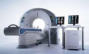
Technology is helping doctors take a 3-D look at the hearts of possible heart attack victims life-saving. High-tech cardiac scans can provide three-dimensional images of the heart’s anatomy and blood circulation. It’s a non-invasive test and can detect if the heart’s blood vessels are blocked or narrowed.

The scan, the 64-slice CT scanner, is a huge advancement in cardiology and uses a combination of X-ray and computer technology. The life-saving machine produces cross-sectional images, often called “slices”, of the heart. The 64-slice scanner can spot things that couldn’t be seen on older scanners, such as the narrowing of arteries that cause heart attacks.
Low levels of radiation are used to create the image, so there is a risk of radiation exposure which may lead to cancer.

